BONE SCINTIGRAPHY
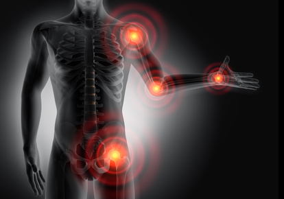
The sceleton scintigraphy, also known as bone scintigraphy, is an imaging method of nuclear medicine. The bone scintigraphy is used as verification of bone parts with heightened bone metabolism.

Areas with heightened bone metabolism – also called focus – are found in bone metastases (e.g. prostate cancer, breast cancer), as well as in healing areas of bone fractures or at inflammatory transformations such as osteomyelitis, inflammatory joint deceases or joint loosening (e.g. hip joint or knee joint prosthesis).
When is this examination necessary?
If you suffer from obscure bone and/or joint pain, this examination method gives information about pathologically changed bone metabolism.
In our practice, we use the bone scintigraphy especially as a preliminary examination for radiosynoviorthesis (RSO).
Process
A radioactive substance (99mTc-HDP) is injected into the bloodstream via arm vein. Within hours, this substance slowly attaches itself to the skeletal structure.
Within the first hour after injection, patients must drink about 1l of liquid to improve the imaging quality. Please make sure to empty your bladder frequently, because the substance is released through the kidneys.
The radiation exposure is like that of a computer tomography.
Please make sure to bring a beach towel to your appointment.
- Loosening of joint prosthesis
- Inflammatory joint diseases
- Search for metastases (e.g. prostate cancer, breast cancer)