
 Raiffeisenstr. 4, 74360 Ilsfeld
RADIOSYNOVIORTHESIS
THERAPY OF INFLAMMATORY JOINT DISEASES
With inflammatory joint diseases the synovium is permanently inflamed. Trigger respectively cause could be - among other things - intense strain or laparoscopic intervention.
If the inflammatory process isn’t stopped in time, damage on chondral and bone will occur, and can even destroy them.
With radiosynoviorthesis (short RSO) these inflammatory joint diseases can be treated.
The therapy is handled on an out-patient basis and scantly painful. If necessary, multiple joints can be treated at the same time.
Depending on the size of the joint, the radio nuclide is chosen (e.g. when treating a finger joint, Erbium is used).
There are three types of radio nuclide - so-called beta emitter - for therapy at hand. The range of radiation is extremely low and only takes effect inside the treated joint. The adjoining tissue usually isn’t reached, meaning there’s no damage to be expected. The radiation exposure for your surroundings is due to the low range of radiation also quite small, which is why RSO can be performed on an out-patient basis.
THE NUCLIDES AND THEIR USAGE:
Yttrium-90: knee joints
Rhenium-186: shoulder, elbow, hand, hip and ankle joints
Erbium-169: finger and toe joints
How do I know if the radiosynoviorthesis is suitable for me?
By means of a 3-phase scintigraphy it is tested if a radiosynoviorthesis is promising in your case.
The scintigraphy is a highly sensitive method for verifying inflammatory processes inside joints as well as changes of osseous areas.
At the beginning of the examination you will be injected with a radioactive substance (99mTc Pertechnetat) via arm vein. It will concentrate itself at the inflamed joints.
The first image performed with the gamma cam is carried out a few minutes after injection. After 2 to 3 hours another image is taken. Another injection isn’t needed.
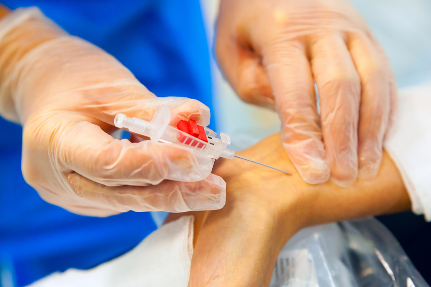
How is the radiosynoviorthesis performed?
The treatment is performed under sterile conditions and under roentgenoscopy (X-ray). The joint puncture will be performed with a thin needle via which a small amount of a local anaesthetic is applied, followed by a contrast medium to revise the correct position of the needle.
After revising the needle’s position, a radioactive substance is applied into the joint, followed by a cortisone injection.
Depending on the treated joint, a scintigram to monitor correct distribution of the activity is performed.
After that a bandage is applied on the treated joint to ensure the necessary immobilization. The bandage can be removed after 48 hours.